Back Of Skull Anatomy : Crossfit The Bones Of The Skull / The occipital bone is located on the back of the cranium and includes.. Inside the skull, it forms the anterior cranial fossa, which contains the frontal lobes of the cerebrum. The occipital bone is located on the back of the cranium and includes. Upon reaching maturity, our skull bones fuse to produce a rigid protective shell for the soft nervous. The bbc is not responsible for the content of external websites. It offers protection to the brain, eye balls, inner ears, and nasal passages.
This anatomic region is complex and poses surgical challenges for otolaryngologists and neurosurgeons alike. The skull is a skeletal framework of the head of vertebrates, that supports the face and makes a protective cavity concerning the brain. It is comprised of many bones, formed by intramembranous ossification, which are joined together by sutures (fibrous joints). The skull performs vital functions. The skull bones can be classified into two groups:
Back Of Skull Bones Diagram Quizlet from o.quizlet.com During childhood development, the skull bones remain somewhat separated, allowing for growth of the brain and skull. The anterior fossa is formed by the orbital plates of the frontal bone, cribriform plate of the ethmoid, and lesser wings of the sphenoid. • it has the supraorbital foramen, where the supraorbital the paired parietal bones make up the top and lateral aspects of the cranium. Skull, skeletal framework of the head of vertebrates, composed of bones or cartilage, which form a unit that protects the brain and some sense organs. This anatomic region is complex and poses surgical challenges for otolaryngologists and neurosurgeons alike. Excluding ear ossicles, it is made of 22 bones. The cranium and the mandible. These joints fuse together in adulthood.
Anatomy next provides anatomy learning tools for students and teachers.
The occipital bone is located on the back of the cranium and includes. 12 photos of the bone of back of skull. Anatomy & physiology · anatomy and physiology. Human skull from the front. • it has the supraorbital foramen, where the supraorbital the paired parietal bones make up the top and lateral aspects of the cranium. In order to be light, the skull is made up by flat and irregular bones, and has hollow spaces called the sinuses. These joints fuse together in adulthood. Excluding ear ossicles, it is made of 22 bones. The skull includes the upper jaw and the cranium. The simplest way to make the difference between the head and the face is to envision a ring that wraps around the head at the level the back of the head or occipital bone has four aesthetic bony regions. The skull is a bony structure that supports the face and forms a protective cavity for the brain. It is comprised of many bones, formed by intramembranous ossification, which are joined together by sutures (fibrous joints). From an anatomical perspective, the skull is divided into two parts:
During childhood development, the skull bones remain somewhat separated, allowing for growth of the brain and skull. The axial & appendicular skeleton. Overview, anterior skull base, middle skull base march 18, 2017. In order to be light, the skull is made up by flat and irregular bones, and has hollow spaces called the sinuses. The anterior fossa is formed by the orbital plates of the frontal bone, cribriform plate of the ethmoid, and lesser wings of the sphenoid.
The Skull Boundless Anatomy And Physiology from s3-us-west-2.amazonaws.com The frontal, parietal, temporal and occipital bones are joined at the cranial sutures. The skull base is the inferior portion of the neurocranium. The bbc is not responsible for the content of external websites. Cranium) is the skeleton of the head composed of 22 separate bones joined together primarily by sutures. They don't move and united into a single unit. The axial & appendicular skeleton. It supports and protects the face and the brain. The skull is the bony skeleton of the head.
These joints fuse together in adulthood.
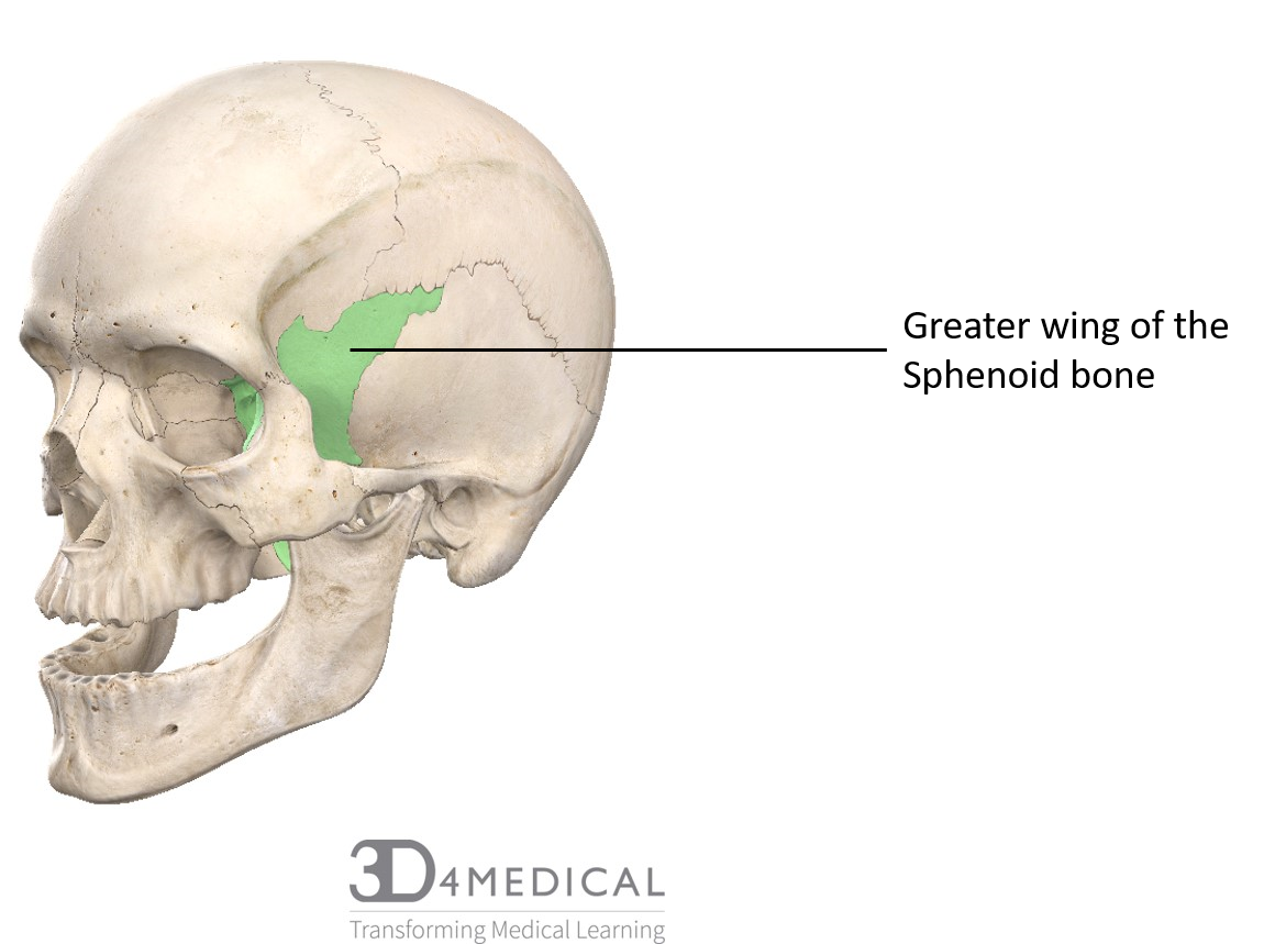
The skull is the bony skeleton of the head. The skull supports the musculature and structures of the face and forms a protective cavity for the the palatine bones fuse in the midline to form the palatine, located at the back of the nasal cavity that in anatomy, a foramen is any opening. Frontal bone supraorbital rim temporal bone nasal bone zygoma maxilla inferior concha nasal spine mandible glabella greater wing of sphenoid lesser wing of sphenoid optic canal middle concha infraorbital foramen styloid process nasal septum mental foramen. The bbc is not responsible for the content of external websites. Learn more about the anatomy and function of the skull in humans and other vertebrates. The major sutures are the coronal suture, sagittal suture, lambdoid suture and squamosal sutures. Excluding ear ossicles, it is made of 22 bones. Looking at it from the inside it can be subdivided into. Human skull from the front. It is comprised of many bones, formed by intramembranous ossification, which are joined together by sutures (fibrous joints). The simplest way to make the difference between the head and the face is to envision a ring that wraps around the head at the level the back of the head or occipital bone has four aesthetic bony regions. Anatomy & physiology · anatomy and physiology. The skull begins to form prior to week 12 of embryogenesis.
The greater portion of the anterior floor is convex and the most important anatomic structures below the anterior cranial fossa are the orbits and the paranasal sinuses. The skull is a bony structure that supports the face and forms a protective cavity for the brain. Axial muscles of the head, neck, and back. The skull base is the inferior portion of the neurocranium. A thorough description is beyond the.
Bones Advanced Anatomy 2nd Ed from pressbooks.bccampus.ca Human skull from the front. The base of the skull (or skull base) forms the floor of the cranial cavity and separates the brain from the structures of the neck and face. During childhood development, the skull bones remain somewhat separated, allowing for growth of the brain and skull. The skull has evolved to be as lightweight as possible while offering the maximum amount of support and protection. The skull performs vital functions. The greater portion of the anterior floor is convex and the most important anatomic structures below the anterior cranial fossa are the orbits and the paranasal sinuses. It offers protection to the brain, eye balls, inner ears, and nasal passages. Inside the skull, it forms the anterior cranial fossa, which contains the frontal lobes of the cerebrum.
The skull includes the upper jaw and the cranium.
The occipital bone is located on the back of the cranium and includes. Foramina inside the body of humans and other animals. The skull bones can be classified into two groups: A thorough description is beyond the. The skull is a bony structure that supports the face and forms a protective cavity for the brain. Skull bones aren't fused together at birth. It offers protection to the brain, eye balls, inner ears, and nasal passages. So, the human skull consists of 23 bones. Skull reshaping is done on any of the structures that lie above the face. Human skull from the front. Inside the skull, it forms the anterior cranial fossa, which contains the frontal lobes of the cerebrum. The skull includes the upper jaw and the cranium. This anatomic region is complex and poses surgical challenges for otolaryngologists and neurosurgeons alike.


0 Komentar